ASTRA beamline
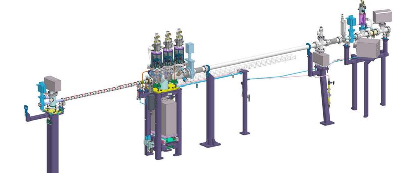
ASTRA (formerly known as SOLABS) is the new beamline at the SOLARIS Centre, which has officially opened in July 2022. The primary analytical technique available at the beamline is X-ray absorption spectroscopy (EXAFS: Extended X-ray Absorption Fine Structure and XANES: X-ray Absorption Near Edge Structure). The beamline uses radiation from the bending magnet, in the energy range of 1keV – 15keV, which covers K absorption edges of the chemical elements between Mg and Se, as well as L and M edges of many other elements. The measurements are possible in ambient pressure, as well as in dedicated reaction cells.
The beamline is the joint project of Hochschule Niederrhein, Synchrotron Light Research Institute, Bonn Uniersity and SOLARIS.
The beamline is optimized for applications in the tender X-ray energy range. The only window between the storage ring and the monochromator is made of a very thin polymer layer, which doesn’t absorb photons in the energy range of 1keV-4keV (which is a common problem for the standard Be or Al windows). The radiation monochromator utilizes not only silicon and germanium crystals, but also indium antimonide, beryllium and multilayer/organic crystal systems, enabling measurements for the photon energies down to 1keV. This energy range covers the K absorption edges of low-Z elements, such as Mg, Al, Si, P, S, making it useful for the life science, pharmaceutical, rubber, chemical, environmental and agricultural industry sectors.
X-ray absorption spectroscopy
X-ray absorption spectroscopy, which will be the main method available at the SOLABS beamline, is based on the absorption of X-ray photons by the studied materials. When the energy of the incident photons approaches the excitation energy of electrons in certain atoms (absorption edge), the absorption coefficient rises significantly. This energy is characteristic for a given element and electronic shell, therefore the method can be used to study the chemical composition of the complex compounds. Thanks to the subtle variation of the absorption coefficient near the absorption edge, which can be probed by varying the energy of the incoming X-ray radiation, chemical properties (such as oxidation state) and local geometric arrangement of the atoms surrounding probed element (distance to the nearest neighbors and their number) can be studied.
In most SR facilities XAS beamlines are the “workhorses” and the most oversubscribed beamlines. For speciation analysis of an element of interest, XAS is the technique of first choice. First results, such as the likely oxidation state of the compounds of the samples, can be easily and directly obtained, using the ‘fingerprint approach’: comparing the spectra of the samples with those of suitable reference compounds. More advanced analyses, requiring computational modeling are also possible, although they require more time and effort.
Even though XAS has been increasingly applied to the various research problems and speciation analyzes from the biological, agricultural, environmental and material sciences, this technique is still not considered a standard method for industrial applications. XAS measurement services, which encompass both the measurement and the interpretation of the obtained data, in order to solve specific R&D problems are currently available only at very few synchrotron light sources in Europe. At the same time, XAS measurements of low Z-elements (down to the Si K-edge) are currently not offered in Europe for commercial users. Since speciation analysis in situ can contribute in many ways to process elucidation and the understanding of chemical reactions, there is great potential for applied research and interest in this technique.
High-resolution spectroscopy
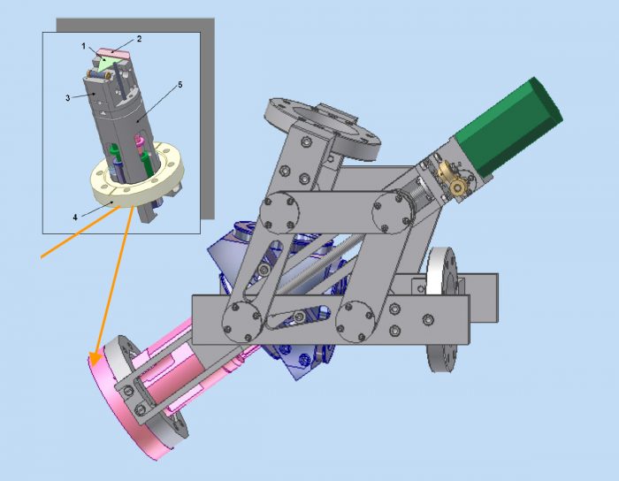
The capabilities of the SOLABS beamline will be enhanced in the Sylinda project by the installation of a new, fluorescence spectrometer, which will enable measuring XAS spectra with very high energy resolution. The spectrometer is based on a design by NIST and it was developed and tested in collaboration between Bonn University and the Karlsruhe Institute of Technology (KIT). In addition to the greatly enhanced resolution, the detector will also allow for the advanced studies, using resonant X-ray emission spectroscopy (REXS), inelastic X-ray scattering (RIXS) and X-ray Raman scattering (XRS) techniques.
The spectrometer is a compact vacuum instrument, enabling measurements for the energies down to 1.7 keV, covering the K-edges of Si, P and S. SOLARIS will be the only synchrotron facility offering this specific technique to external users from industry in Europe. This will make it a favored synchrotron radiation facility for cooperation with commercial entities, including SMEs, especially in case of working with the samples containing low Z-elements, which include biological samples, or answering research questions, requiring high-resolution spectroscopy.
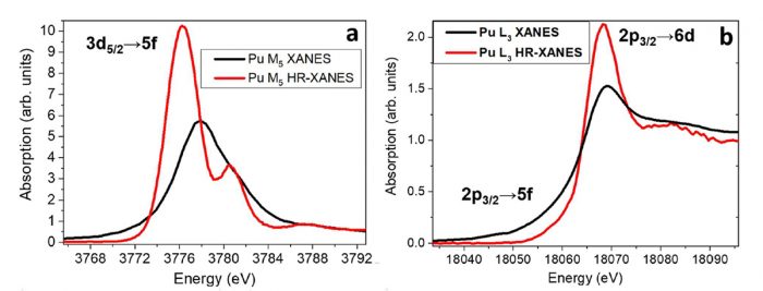
For “standard” XAS experiments when spectra are measured either in transmission mode or in fluorescence mode using an energy dispersive detector, the energy resolution of the spectra is limited by the natural lifetime broadening of the core hole, not the incident beam energy resolution. In the K-shell photoabsorption process, for example, the incident photon ejects the K-shell electron to the unoccupied states, and since this hole has a finite lifetime the hole is filled by an outer-shell electron leading, in the case of the radiative decay, to fluorescence radiation. The Heisenberg uncertainty principle gives rise to an energy uncertainty inversely proportional to the lifetime, for the K shell spectra ranging from about 1 eV (V, Z =23) up to 40 eV (W, Z =74). With high resolution energy analysis of the fluorescence radiation using, for example, a wavelength dispersive crystal spectrometer the lifetime broadening can be overcome. For these experiments normal fluorescence X-ray absorption near edge structure (XANES) spectra are recorded by scanning the incident beam across an absorption edge and monitoring the fluorescence intensity using an analyser with better resolution than the natural lifetime width. Spectra that are recorded using this technique show significantly more details than the “conventional counterparts”. An example, showing the direct comparison of a conventional (black) and a high resolution (red) Pu M5/L-III XANES spectra taken for the same samples is shown below:


